The Cellular and Supramolecular Structure and Function (CSSF) Section of LCIMB is located in Building 13, Room 3E63.
The laboratory has facilities for preparing tissues, cells, and isolated macromolecular assemblies for electron microscopy, either in the CSSF Section or in the neighboring NIH-wide Electron Microscopy Shared Resource. Preparative equipment includes a Baltec HPM10 high-pressure freezing machine, FEI vitrobot for freezing EM grids, Leica UCT/FCS cryo-ultramicrotome, Leica EM UC6 ultramicrotome, Leica EM/AFS2 freeze-substitution system, EMS and Edwards 306 carbon evaporators.
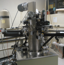
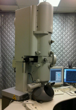
The laboratory’s dedicated electron microscope is an FEI Tecnai TF30 TEM operated at an accelerating voltage of 300 kV and equipped with a field-emission source. The instrument is also equipped with a Gatan Tridiem imaging filter, two 2k x 2k pixel Gatan Ultrascan cooled CCD cameras, a Fischione HAADF detector, Fischione dual-axis tomography holder, Gatan cryo-transfer tomography holder. The instrument is also has FEI and Gatan software packages for performing electron tomography, hyperspectral imaging, electron energy loss spectroscopy, and scanning transmission electron microscopy. Through the neighboring Electron Microscopy Shared Resource, the laboratory also has access to an FEI T12 TEM operating at an accelerating voltage of 120 kV, and equipped for electron tomography, and energy-dispersive x-ray spectroscopy. Also available are a Hitachi H4800 field-emission scanning electron microscope, and a Zeiss Sigma SEM equipped with a Gatan 3View serial blockface imaging system.